


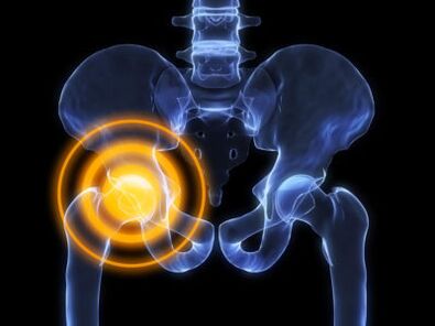
Hip joint arthrosis is a complex disease with specific symptoms and complex treatment.The disease occurs against the backdrop of metabolic disorders in the cartilage tissue of the joint cavity and the femur's head.
Hip or coxartrosis arthritis is more common in the elderly.It is generally acknowledged that an inflammatory reaction plays a major role in the pathogenesis of the disease.After a large number of studies, it was proved that arthritis occurs with atherosclerosis and diseases with metabolic disorders.
The essence of the disease
Coxartrosis is a disease based on metabolic disorders with atrophic and degenerative changes in the cartilage tissue of the hip joint.
You cannot confuse arthritis with arthritis.Unlike arthritis with arthritis, non -infectious (aseptic) inflammation occurs, which develops and progresses for many years.
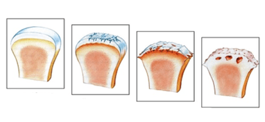
Pathogenesis of development:
- Violation of metabolic processes in the cartilage.Cartilage tissue receives nutrients using diffusion.Inflammation or smaller edema leads to the absence of trace elements and minerals.
- In contrast to the backdrop of the processes damaged by the nutrients, the atrophic changes begin, the cartilage tissue is refined, the amount of joint fluid and the chondroblasts decreases.
- Due to the folly and destruction of the cartilage, severe pain begins, there is a decrease in amplitude of movements in the joints.
- The cartilage fabric is very sophisticated, the fabulous cleft is narrowing, the common dystrophy occurs.
Until the dystrophy begins, for more than a year it develops.The disease can only stop at the early stages, with the development of the third phase of osteoarthrosis, the treatment is aimed at reducing the symptoms and claiming the patient's life, an alternative to medication - endoprosthetics.
Reason
The disease is poletiological, there are many conditions and factors that can lead to arthritis or provoke its progress.If the causes of hip joint arthrosis are not detected, such a disease is called idiopathic arthrosis.
The disease is not hereditary, but the genetic pathologies in which the cartilage dysplasia occurs can cause arthrosis of the hip joint.
Also, the cause of coxarthrosis can be such diseases:
- APLE Syndrome - a characteristic sign of the disease is a violation of the distribution of nutrients in the cartilage tissue of the femur and the head of the femur.It happens in childhood, mostly boys are sick.
- Congenital displacements and femur subluxation.In the process of damage, an inflammatory reaction and aseptic melting of the cartilage tissue and the femoral head may occur.
- Necrosis of the femur's head.It occurs due to damage to the upper artery, which is glued to the top of the head.
- Rheumatoid and juvenile arthritis.In contrast to the backdrop of the action of its toxins or antibodies, exudative inflammation develops in the joint.
Given the fact that the disease progresses slowly, the disease can be on one side and two.
There are many factors that contribute to the appearance of arthrosis, they include:
- Spinal diseases (kyphosis, lordosis, scoliosis);
- Metabolic diseases of connective tissue;
- violation of blood supply to the joints;
- atherosclerosis of large vessels;
- states of stress;
- hip dysplasia;
- congenital deformities of the lower extremities;
- Infectious diseases;
- inactive lifestyle;
- alcohol intake, smoking;
- old age.
Remember that people who are engaged in stretching have a great risk of developing arthrosis at adulthood.
Also, one of the reasons may be the traumatic damage to the components of the union.After tissue damage occurs, an inflammatory reaction occurs, as a result of which the cartilage can be replaced with the connector.
Symptoms
Due to the fact that the disease is progressing slowly, the patient does not always pay attention to the first signs.It should be noted that with the early diagnosis, the chances of remission of the disease increase.It is very important to start treatment earlier, as it is possible to avoid the appearance of ankylosis and complete osteoarthrosis.
With hip joint arthrosis, symptoms can occur with different intensities depending on the loads and the degree of the disease.
Clinical photography of hip union osteoarthritis:
- Painful sensations that grow in severe pain in the front and lateral thigh.Patients complain that the thigh hurts a lot during the turn or load on the joint.
- The unpleasant sensations that arise in the groin when walking, sometimes they combine with pain in the thighs.
- The stiffness and limitation of limb motility in the hip joint.First, the removal function suffers, and then everyone else.
- Uncomfortable sounds when walking, the knot can click or cramp.Constant pathological sounds can be the only sign of the disease.
- Morning rigidity, which passes within two hours or before dinner.
Sometimes ignoring the possible consequences, people begin to take medication for symptomatic therapy and thereby mask the progress of the process of destruction in the cartilage.
Degree of illness
Clinical photography depends on the degree of hip joint arthrosis and the reactivity of the patient's body.If symptoms appear, as a rule, changes occur in X -rays.In medical practice, it is common to distinguish three radiological stages, each of which has its own characteristics.
The degree of arthrosis in relation to changes in X -Rids:
First degree arthrosis
It continues with minimal clinical manifestations, and therefore patients rarely seek help from a doctor.With an early diagnosis of the disease, the patient increases the chances of complete recovery.The initial period of the disease is characterized by minor pain in the pelvis and thigh, the pain increases against the backdrop of physical exercise or prolonged walking.In the second place in terms of frequency of manifestation comes a symptom of pain in the groin.At 1 degree, the pain is withdrawing and rarely happening.The volume of movements is fully preserved.In an X -Ray, small changes are visualized.
Second rank
In case of a second degree, the patient begins to disturb more acute and frequent pain that may occur at rest.Symptoms, as a rule, manifest in the evening hours and breakfast stiffness does not pass until dinner.During prolonged transitions, a symptom of dementia occurs, a person cannot fully charge the diseased joint.The discomfort occurs during flexion or squats, degenerative processes progress to the cartilage.Against the backdrop of such changes, the foot can shorten, the atrophy of the hip and pelvis muscle occurs.In radiography, a narrowing of the joint gap is visible, a periostal reaction increases.A large number of osteophytes are found in the lumen of union.
3rd stage of final or scattered arthrosis
The third stage is characterized by the appearance of motor dysfunction of the lower extremities.The patient complains of persistent pain, which does not occur for no reason.There is an abbreviation of the limbs more than 5%, ankilosis occurs, the union loses the ability to mobility.Radiography indicates complete closure of the common gap and a large number of osteophytes against the background of bone deformation.3 -degree treatment is performed only in an operational way.
Treatment methods
The choice of methodology for treatment depends on the degree of osteoarthritis.In the early stages, comprehensive conservative treatment is used.The most difficult is the second phase, as conservative therapy is ineffective, and indications for surgery are not enough.Completely is completely possible to cure arthritis only with the development of the first degree of disease.
After making a diagnosis of hip joint arthrosis, the doctor chooses treatment methods.Most used:
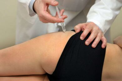
- conservative treatment with medication;
- surgical treatment;
- Therapy and massage of exercises.
Each of the treatment methods has its own characteristics, changes and specific goals.Conservative therapy is used for such purposes:
- The fight against the etiological factor.For example, metabolic or hormonal disorders can be corrected.
- Symptomatic treatment that aims to facilitate the patient's life and relieve the symptoms of aliteration.For this purpose, non -steroidal anti -inflammatory drugs are used.Most often, diclofenaci sodium, nimesulide, ibuprofen are used by NSAIDs.
To get rid of persistent pain, NSAIDs are taken almost daily, and this can affect the patient's gastrointestinal tract and cause the development of peptic ulcer.
Surgical intervention is shown with the third degree of arthrosis and is the only method for restoring the function in the foot.The essence of the methodology is complete or partial replacement of the joints of the joint with titanium endoprostees.
Physical media education is an integral part of any measure of rehabilitation.Exercise therapy and massage are aimed at improving the blood flow to the joint.Also, exercise therapy is used to reduce the risk of ankylosis.
You need to be careful when doing exercises, as you can damage the common osteophytes.Tactics and exercises should be chosen by a physician based on your individual characteristics and clinical appearance of the disease.